מערכת ממוגרפיה מתקדמת עם Tomosynthesis
נגישות
גודל טקסט
רגיל
גדול
ענק
תצוגת צבעי האתר (* פועל בדפדפנים מתקדמים מסוג Chrome ו- Firefox) תצוגה רגילה מותאם לעיוורי צבעים מותאם לכבדי ראייה
עיצוב האתר עיצוב רגיל תוכן בלבד, ללא עיצוב
סגור
תצוגת צבעי האתר (* פועל בדפדפנים מתקדמים מסוג Chrome ו- Firefox) תצוגה רגילה מותאם לעיוורי צבעים מותאם לכבדי ראייה
עיצוב האתר עיצוב רגיל תוכן בלבד, ללא עיצוב
סגור

מערכות ממוגרפיה
gr
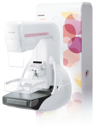
AMULET Innovality
וידיאו

Innovation and quality in mammography. The new leader in the AMULET series. Tomosynthesis, 3D mammography and biopsy are all available
Features
Unique new detector for fast, low dose examinations
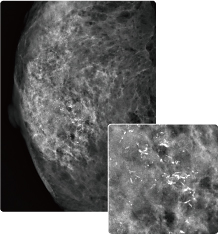
AMULET Innovality employs a direct-conversion flat panel detector made of Amorphous Selenium (a-Se) which exhibits excellent conversion efficiency in the mammographic X-ray spectrum. The new HCP (Hexagonal Close Pattern) detector efficiently collects electrical signal converted from X-rays to realize both high resolution and low noise. This unique design makes it possible to realize a higher DQE (Detective Quantum Efficiency) than with the square pixel array of conventional TFT panels. With the information collected by the HCP detector, AMULET Innovality creates high definition images with a pixel size of 50 μm; the finest available with a direct-conversion detector.
To simplify the general radiography examinations required for various techniques, the ceiling type X-ray tube support features excellent operability and incorporates an LCD touchpanel to set both the exposure parameters and Anatomical Programs (APR).In combination with Bucky table interlock functions, these features reduce the labor required for examinations.The system provides a comfortable examination environment for operator and patient alike.
This low-noise and high-speed switching technology allows tomosynthesis exposures with a low X-ray dosage and short acquisition time to be performed. Fast image display is also possible, realizing a smooth mammography aworkflow from exposure to image display.
 |
 |
| Conventional square pixel | AMULET Innovality hexagonal pixel |
|---|
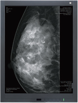
Tomosynthesis: making it possible to observe the internal structure of the breast
|
In breast tomosynthesis, the X-ray tube moves through an arc while acquiring a series of low-dose x-ray images. The images taken from different angles are reconstructed into a range of Tomosynthesis slices where the structure of interest is always in focus. The reconstructed tomographic images make it easier to identify lesions which might be difficult to visualize in routine mammography because of the presence of overlapping breast structures. |
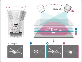 |
The Tomosynthesis function on AMULET Innovality is suitable for a wide range of uses, offering two modes to cater for various clinical scenarios. Standard (ST) mode combines rapid exposure timing and efficient workflow with a low X-ray dose while High Resolution (HR) mode makes it possible to produce images with an even higher level of detail, allowing the region of interest to be brought into clearer focus.
Two modes suitable for a range of clinical purposes
ST (Standard) modeAcquisition angle: ±7.5° Pixel size: 150/100 μm The smaller angular range and fast image acquisition allow Tomosynthesis scans to be quickly performed with a relatively low x-ray dose. |
 |
HR (High Resolution) modeAcquisition angle: ±20° Pixel size: 100/50 μm With a larger acquisition angle the depth resolution is improved. This allows the region of interest to be defined more clearly and brought into clearer focus. |
 |
 |
 |
Intelligent AEC optimizes the X-ray dose for each breast type
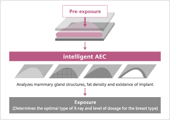
Intelligent AEC has advantages in defining the optimal dose for an examination compared to conventional AEC systems where the sensor position is fixed. Through the analysis of information obtained from low-dose preshot images, Intelligent AEC makes it possible to consider the mammary gland density (breast type) when defining the x-ray energy and level of dose required. Able to be used even in the presence of implants; intelligent AEC enables more accurate calculation of exposure parameters than is possible with conventional AEC systems. By allowing the use of automatic exposure for the implanted breast, Intelligent AEC can further enhance examination workflow.
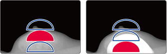
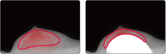
Conventional AEC |
Intelligent AEC |
 |
 |
|
Requires manual adjustment of the settings based on the assumed location of mammary gland |
Automatically selects the region for exposure in the pre-shot image |
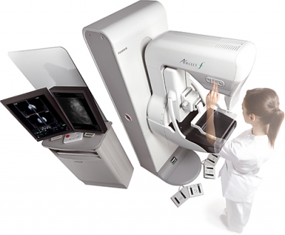
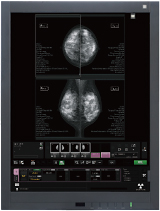
Dedicated mammography AWS (Acquisition Workstation)
Optimal examination workflow
|
 |
 |
| AWS | High definition second monitor |
High definition second monitor (3M/5M: Optional)
- A second, high resolution monitor can be added to the AWS making it possible to display previous images recalled from a PACS to ensure the mammographer has access to previous images at all times.
- For Tomosynthesis, reconstructed images can be displayed and subjected to image QC.
מוצרים מקושרים
מערכת הממוגרפיה הדיגיטאלית המתקדמת ביותר בעולם
פרטים נוספיםAmulet f
אודות דינקו
כבר מעל ל-40 שנה נמצאת דינקו בחזית טכנולוגיות הדימות הרפואיות והתעשייתיות בישראל, כספק מוביל של ציוד ומערכות מידע רדיולוגיים.
חברת דינקו הינה הנציגה הבלעדית בישראל של החטיבות הרפואיות של ענקיות הרפואה FUJIFILM ו-Shimadzu. צוותי השירות והפיתוח שלנו תומכים בכל הארגונים הרפואיים בישראל ובגופים המובילים בתעשיות האזרחיות והבטחוניות.
יצירת קשר
דוא"ל: This e-mail address is being protected from spambots. You need JavaScript enabled to view it.
טלפון: 04-8131515
פקס: 04-8131516