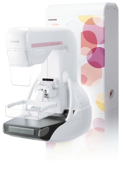
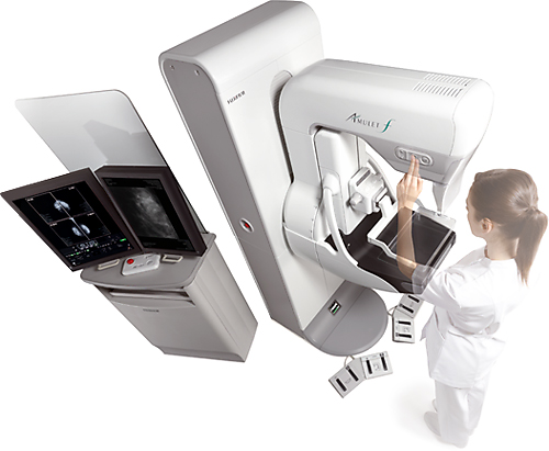
מערכת ממוגרפיה דיגיטאלית בעלת איכות תמונה בלתי מתפשרת, נוחות ויעילות!
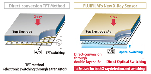
איכות זו מתקבלת בעזרת החיישן הייחודי של Fuji, מסוג Direct Optical Switching Technology המשלב חיישן מסוג Direct Conversion Flat Panel Detector. החיישן בטכנולוגיה זו כולל שכבה כפולה של Amorphous Silicone המספקת למערכת רגישות לרנטגן ואיכות תמונה גבוהים במיוחד - שילוב אידיאלי בזיהוי מיקרוקלסיפיקציות וגידולים בפירוט רב יותר ולאבחון מוקדם של סרטן השד.
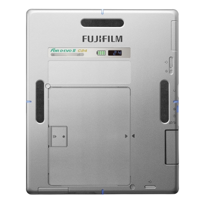
- גודל הפיקסל הקטן ביותר - 50 µm.
- איכות תמונה מצוינת.
- חיישן מסוג Direct Conversion FPD הראשון בעולם.
- חווית ממוגרפיה עדינה ונוחה למטופלת, המפחיתה את חוסר הנוחות בביצוע הבדיקה.
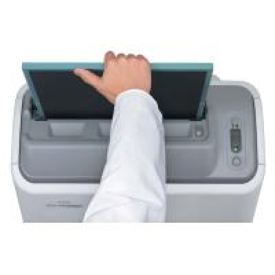
- תהליך בדיקה אופטימאלי, בשימוש עמדת רכישת התמונה AWS Acquisition Workstation.


 The buttons are shaped to be easily identified by touch alone.
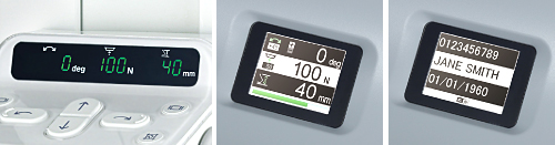
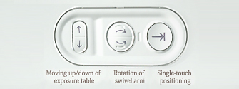
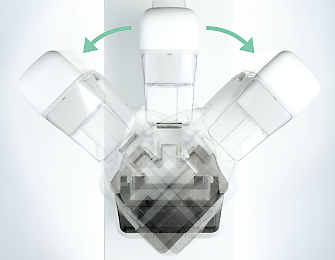 A single-touch function allows the AMULET to be positioned automatically to the desired swivel arm angle between exposures for faster examinations and also returns the unit to the upright position once these are completed.
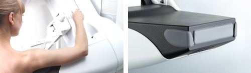
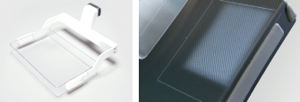 A full range of compression plates are available, including those optimised for small breasts, for more accurate positioning. Both collimation and image output sizes are automatically adjusted to 18×24cm or 24×30cm as required. *Optional
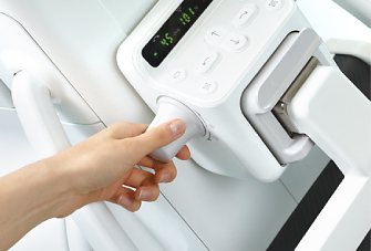 Easy to use controls on the compression arm are provided to allow adjustment of the height of the AMULET and the swivel arm angle, and there is also the ability to vary the automatically set compression force, if necessary, using a simple dial.
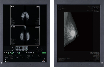 A second high resolution monitor can be added to the AWS making it possible, if connected to PACS, to display previous images to enable more accurate examinations.
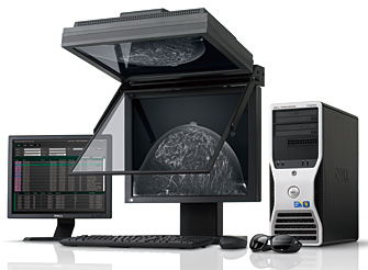 Fujifilm's 3D mammography creates 3D images by using two high resolution images taken from different angles. One of these images is a conventional 2D image. The images are presented on a special viewer. 3D images enable the internal anatomical breast structures to be identified more clearly than in a 2D image due to tissue separation and microcalcifications stratification. With this system, it is expected that image interpretation is as quick or even quicker than 2D mammography and false positives are reduced.
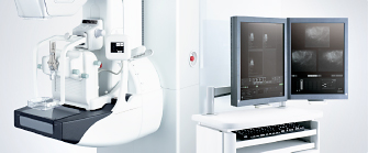 The AMULET's ability to deliver detailed, 50 µm resolution, images to its high resolution display enables precise and efficient biopsy examinations.
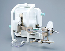 By attaching the lateral adapter, puncture can be performed not only vertically but also laterally to the compressed breast. Two puncture directions are now available, enabling wider application of mammographic needle biopsy.